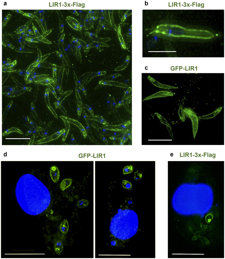
Fig 2. LIR1 localizes at the plasma membrane of promastigotes and intracellular amastigotes.
(a,b) Immunofluorescence deconvolution images of L. amazonensis promastigotes expressing LIR1 with a 3xFlag at the C-terminus. Blue, DAPI DNA staining. Green, anti-3xFlag antibodies. Scale bars: 7 μm (a) and 3.5 μm (b). (c) Deconvolution fluorescence microscopy image of L. amazonensis promastigotes expressing LIR1 fused to GFP at the N-terminus. Scale bar: 11 μm. (d,e) Immunofluorescence deconvolution images of BMM infected for 48 h with axenic amastigotes expressing LIR1 fused to GFP at the N-terminus (d) or 3xFlag at the C-terminus (e). Blue, DAPI DNA staining. Green, GFP-LIR1 or anti-3xFlag antibodies. Scale bars: 11 μm.

