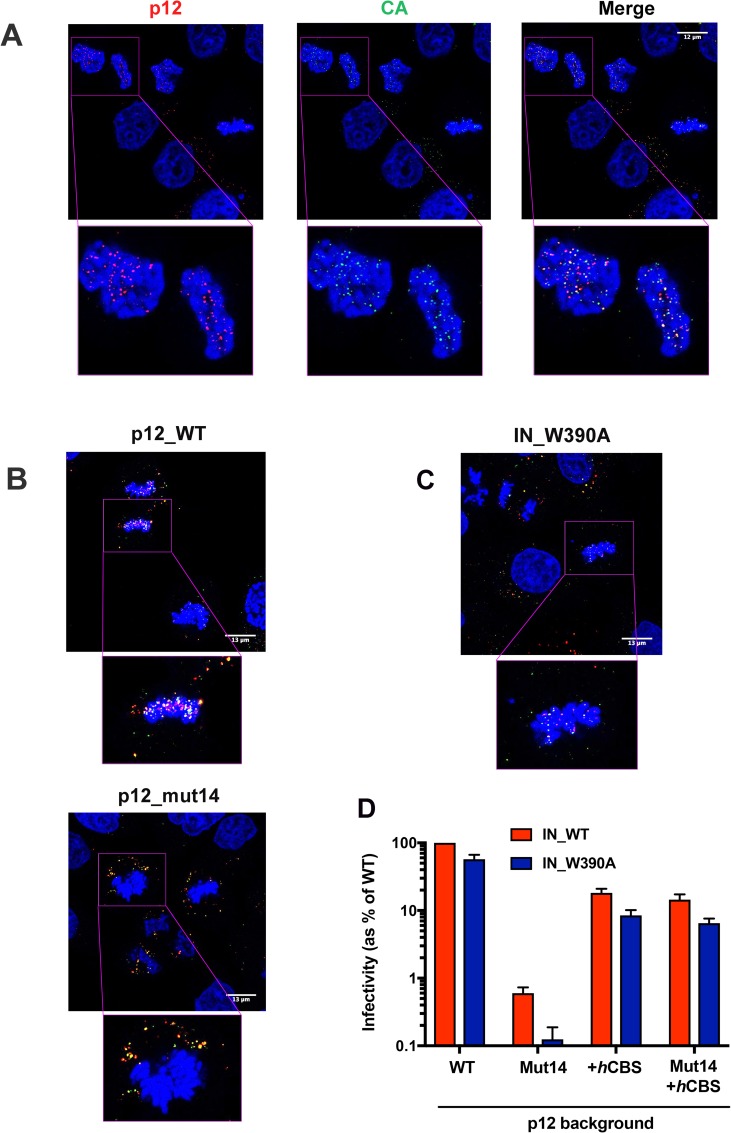
Fig 2. CA and p12 co-localise on mitotic chromatin independently of BET-protein binding.
HeLa cells synchronised using a double-aphidicolin block were infected with WT Mo-MLV VLPs or mutants carrying p12_mut14 or IN_W390A. 10 h post-infection, the cells were fixed, stained for p12 (anti-p12, red), CA (anti-CA, green) and DNA (DAPI, blue), and analysed by confocal microscopy. (A) Representative images showing WT p12 and CA co-localisation on mitotic chromatin. Bottom panels are enlarged views of boxed regions in top panels. (B) Representative images of cells infected with VLPs carrying p12_WT (top panels) or p12_mut14 (bottom panels). (C) A representative image of cells infected with VLPs carrying IN_W390A, which is deficient in BET protein binding. (D) Infectivity of Mo-MLV VLPs carrying alterations in p12 and/or IN. HeLa cells were challenged with equivalent RT units of LacZ-encoding VLPs carrying p12_WT, p12_mut14, p12+hCBS or (p12+hCBS)_mut14 in combination with IN_WT or BET-binding deficient IN_W390A. Infectivity was measured 72 h post-infection by detection of beta-galactosidase activity in a chemiluminescent reporter assay. The data are plotted as percentage of WT VLP infectivity (mean ± SEM of >3 biological replicates).

