
Fig. 1. Differentiation of LY podocytes.
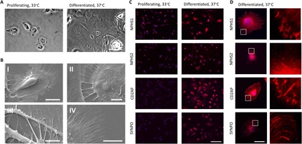
(A) Phase-contrast microscopy of proliferating LY human podocytes cultured at 33°C. Differentiation of LY podocytes at 37°C for 12 days results in a transition to differentiated morphology. Differentiated cells increase in size up to lengths of 150 μm and develop finger-like foot processes around the periphery of the cell. Scale bar, 50 μm. (B) Scanning electron microscopy reveals ultrastructures of primary and secondary foot processes on differentiated LY podocytes. (I) LY podocyte differentiated at 37°C for 12 days demonstrates primary foot processes around the cell periphery and extending toward adjacent cell. Scale bar, 20 μm. (II) Primary foot processes interdigitating between differentiated LY podocytes. Scale bar, 5 μm. (III) Primary foot processes branching off of a differentiated LY podocyte. Scale bar, 5 μm. (IV) Secondary foot processes around the outer periphery of a differentiated podocyte. Scale bar, 10 μm. (C) Localization and expression of podocyte-specific proteins NPHS1, NPHS2, CD2AP, and SYNPO (red) in proliferating and differentiated LY podocytes assessed by immunocytochemistry at low magnification. DAPI (4′,6-diamidino-2-phenylindole) (blue) detects nuclei. Scale bar, 200 μm. (D) Localization and expression of podocyte-specific proteins NPHS1, NPHS2, CD2AP, and SYNPO (red) in a single differentiated LY podocyte. DAPI (blue) detects nuclei. Scale bar, 50 μm; right panels, magnification of area marked by white boxes.