
Fig. 4. Representative posttreatment histology images from a subset of nonhuman primate tissues.
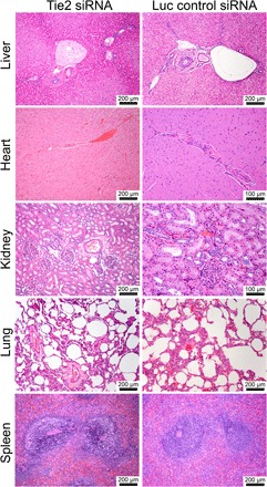
Hematoxylin and eosin were used to stain the tissue. No deleterious effects, such as hepatocellular and cholestatic injury, were observed at this day 2 time point.