. 2018 May 10;7:e36620. doi: 10.7554/eLife.36620

Figure 2. SLC37A3 and ATRAID are functionally related genes required for the mechanism of action of N-BPs.

(A–D) Dose response curves of wild-type, ATRAIDKO and SLC37A3KO K562 cells (A) and HEK 293 T cells (B), and wild-type and SLC37A3KO RAW cells (both undifferentiated macrophages, (C), and differentiated osteoclasts, (D) to alendronate. Cells were treated with a series of doses of alendronate (x-axis) for 48 hr. Relative cell viability was determined by measuring post-treatment total cellular ATP levels and normalizing to those in untreated cells (y-axis). Data depict mean with s.d. for biological triplicate measurements. (E) Immunoblots measuring alendronate-induced reduction in protein prenylation in wild-type and knockout K562 and HEK 293 T cells. Cells were treated with indicated doses of alendronate for 24 hr before analysis by immunoblotting. (F) Immunoblots comparing alendronate-induced reduction in protein prenylation in single and double-knockout HEK 293 T cells. Experimental procedure is as in (C). Note that higher alendronate doses were used in (F) compared to (E) to induce detectable levels of unprenylated proteins. ALN: alendronate.

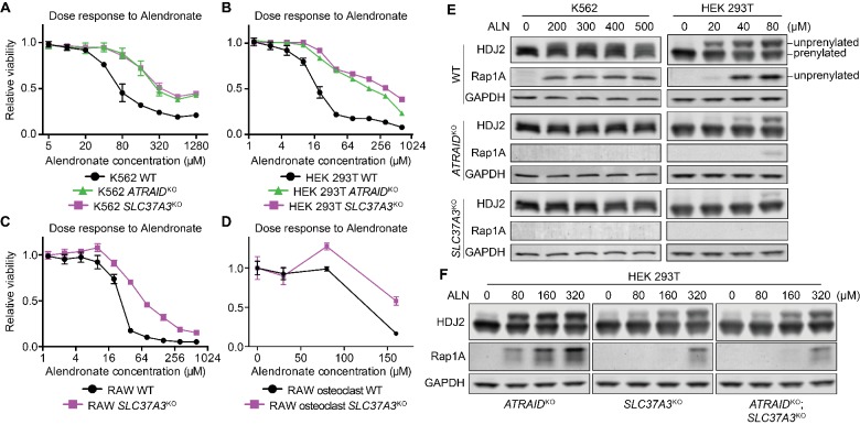
Figure 2.

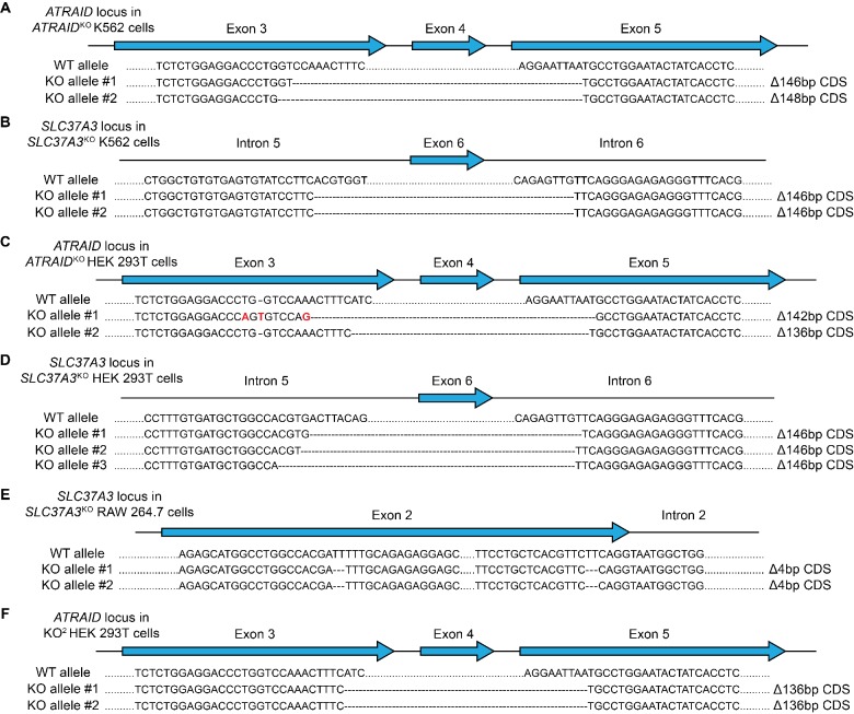
Figure 2—figure supplement 1. Genotypes of knockout cells used in this study.

Figure 2—figure supplement 1.

(A–F) Sequences of the ATRAID (A, C and F) or SLC37A3 locus (B, D and E) in ATRAIDKO (A) and SLC37A3KO (B) K562 cells, ATRAIDKO (C), SLC37A3KO (D) and KO2 (F) HEK 293 T cells, and SLC37A3KO (E) RAW 264.7 cells, showing that truncations in ATRAID and SLC37A3 coding sequences (CDS) have caused frame shifts in the knockout cell lines. The SLC37A3 locus in KO2 HEK 293 T cells is identical to that in SLC37A3KO HEK 293 T cells. Introns and exons are not drawn to scale. Note that more than two alleles are present for the SLC37A3 locus in HEK 293 T cells as these cells are hypo-triploidic. KO2: ATRAIDKO; SLC37A3KO.

Figure 2—figure supplement 2. Additional evidence that validates SLC37A3 and ATRAID as functionally related genes required for the mechanism of action of N-BPs.

Figure 2—figure supplement 2.

(A) Expression of osteoclast markers (Ctsk, Rank and Trap) in undifferentiated RAW macrophages (Mφ) and differentiated RAW osteoclasts (OC), demonstrating successful differentiation of RAW cells. (B–C) Dose responses to alendronate in SLC37A3KO or ATRAIDKO HEK 293 T cells complemented with epitope tagged SLC37A3 or ATRAID, respectively, compared with those in wild-type, ATRAIDKO and SLC37A3KO cells. (D) Immunoblot comparing the short isoform of ATRAID (sATRAID) with the long isoform of ATRAID (lATRAID), showing that the long isoform of ATRAID is expressed as the same protein as the short isoform of ATRAID. Note that unequal amount of protein was loaded in each lane to obtain even exposure. The amount of protein loaded from left to right was: 100 µg, 40 µg, 10 µg and 4 µg, respectively. (E–F) Dose response curves of wild-type, ATRAIDKO and SLC37A3KO HEK 293 T cells to nitrogen-containing bisphosphonates (E) and non-nitrogen-containing bisphosphonates (F). (G–H) Dose response curves of wild-type, ATRAIDKO and SLC37A3KO K562 cells (G) and HEK 293 T cells (H) to lovastatin. (I) Immunoblots measuring lovastatin-induced reduction in protein prenylation in wild-type, ATRAIDKO and SLC37A3KO K562 cells and HEK 293 T cells. (J) Dose response to alendronate in KO2 HEK 293 T cells, compared with those in wild-type, ATRAIDKO and SLC37A3KO cells. For (B–C), (E–H) and (J), data depict mean with s.d. for biological triplicate measurements. LOV: lovastatin. KO2: ATRAIDKO; SLC37A3KO. PNGase F: peptide: N-glycosidase F, an enzyme that removes asparagine (N)-linked sugar modifications on glycoproteins.