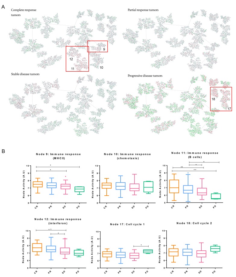
Figure 2. Breast cancer network by pathological response groups.
A. Detail of nodes with the highest activity in each of the subgroups. Genes with an expression below 0 were represented in green; genes with an expression around 0 were represented in grey and genes with an expression above zero were represented in red. B. Functional node activities differences between pathological response groups: Box-and-whisker plots are Tukey boxplots. All p-values were two-sided and p < 0.05 was considered statistically significant. P-value < 0.05 (*); p-value < 0.01(**). A.U: arbitrary units.

