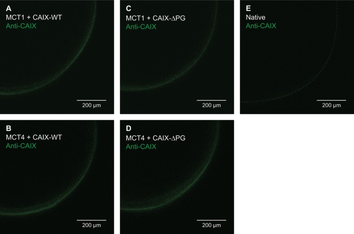
Figure 2. Localization of CAIX-WT and CAIX-ΔPG in Xenopus oocytes.
Antibody staining of fixed and permeabilized oocytes, expressing MCT1+CAIX-WT (A), MCT4+CAIX-WT (B), MCT1+CAIX-ΔPG (C), MCT4+CAIX-ΔPG (D), and a native oocyte as control (E). CAIX was labeled with an antibody, mapping against the C-terminal region of CAIX. Pictures were taken with a confocal laser scanning microscope.

