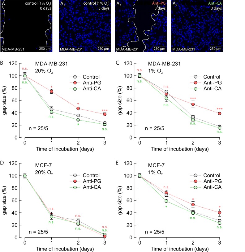
Figure 9. Antibodies directed against the PG domain, but not against the catalytic domain of CAIX inhibit migration of MDA-MB-231 and MCF-7 cells.
(A) Staining of nuclei with Hoechst (blue) in hypoxic MDA-MB-231 cells before (A1) and 3 days after production of a scratch through the culture (A2-A4). Cells remained either untreated (A1, A2) or where incubated with Anti-PG (A3) or Anti-CA (A4). (B-E) Size of the gap (%) in MDA-MB-231 (B, C) and MCF-7 (D, E) cell cultures, 0–3 days after scratching. Cells were incubated under normoxic (B, D) or hypoxic (C, E) conditions in the presence of Anti-PG (red) and Anti-CA (green), respectively, or without antibody (gray). Data are represented as mean ± SEM. Significance in differences was tested with ANOVA, followed by means comparison. The red significance indicators depict differences between cells treated with Anti-PG and control cells, the green significance indicators depict differences between cells treated with Anti-CA and control cells at the respective time points.

