Figure 5.

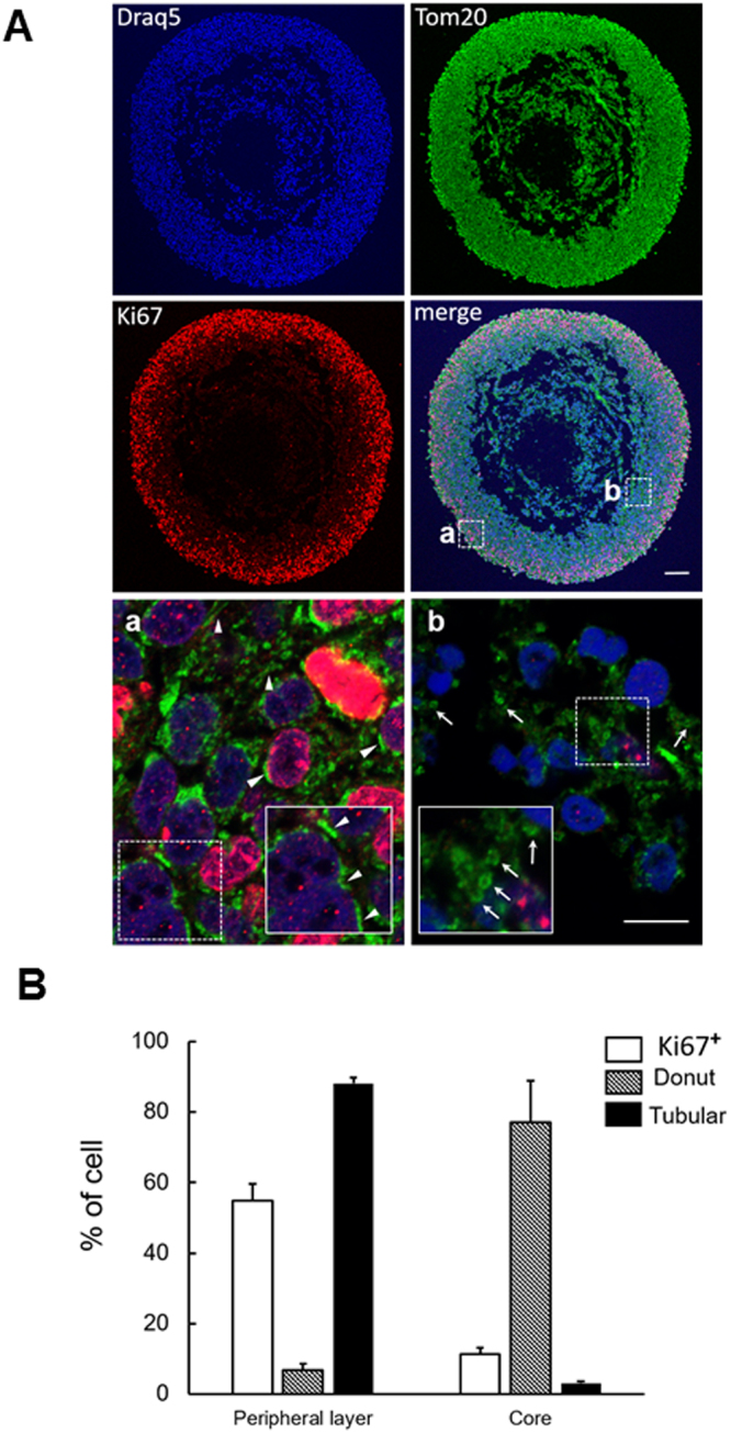
Mitochondrial morphology remodeling in glioblastoma tumorospheres. (A) Confocal microscopy analysis of TG1 macro-tumorosphere section after 8 weeks in culture. Proliferating cells were labeled with the Ki67 antibody (red), mitochondria morphology revealed with the TOM20 antibody (green) and nucleus stained with Draq5 (blue). Arrow-heads point to typical tubular-shaped mitochondria and arrows to typical donut-shaped mitochondria. Insets are enlarged images of selected areas (dotted squares) in (a) and (b) domains of the macro-tumorosphere. Pictures taken with a 10 × 0.30 N.A. objective for whole views of tumorospheres and with a 63X 1.40 N.A. objective for (a) and (b) details on a Leica SP8 upright confocal microscope. Scale bars: 100 µm in all images in A and 5 µm in Aa and Ab. (B) Histogram plot of the number of cells with at least one tubular-shaped mitochondria or one donut-shaped mitochondria in the outer rim (a) and in the core (b) domains of the tumorosphere. 6 areas were counted in each domain.
