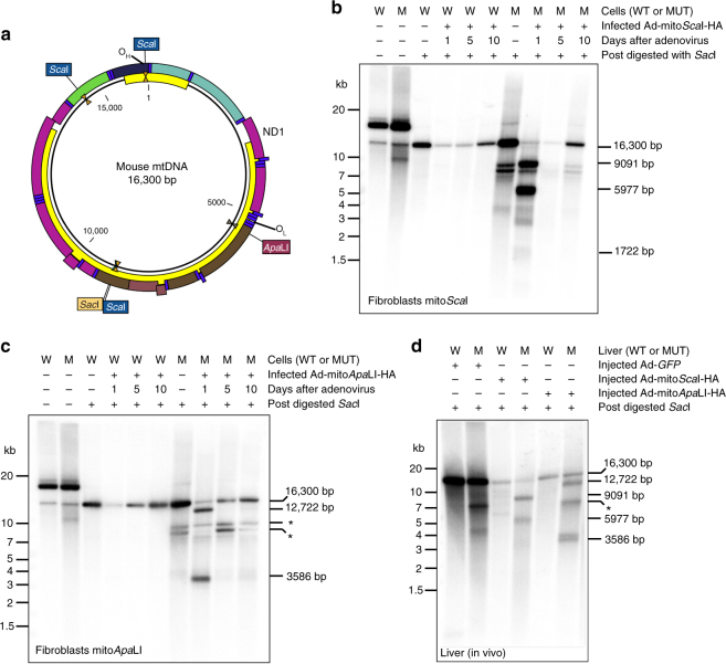
Fig. 2.
Delayed degradation of mtDNA after DSB in DNA from fibroblasts and the liver of the mutator mouse. a Diagram of wild-type C57BL6/J mtDNA illustrating the location and number of restriction endonuclease sites. Southern blot from mutator and wild-type fibroblasts infected with (b) Ad-mitoScaI-HA and (c) Ad-mitoApaLI-HA at 1, 5, and 10 days post-infection. d Southern blot from mutator and wild-type mice liver 5 days after retro-orbital injections with Ad-GFP, Ad-mitoScaI-HA, or Ad-mitoApaLI-HA. DNA was post-digested with SacI to linearize mtDNA. Zeta-Probe membrane was hybridized with a [α-32P] dCTP labeled probe covering 11.7 kb of mtDNA

