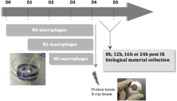
Fig. 1. Schematic outline of the irradiation procedure for macrophages.
THP-1 monocytes were differentiated into macrophages (M0) with 150 nM PMA in cloning cylinder, placed at the center of the irradiation chamber. Macrophages were polarized in M1 phenotype with 10 pg/ml LPS and 20 ng/ml IFN-γ during 24 h incubation or were polarized in M2 phenotype with 20 ng/ml IL-4 and IL-13 during 48 h incubation. THP-1 monocytes were differentiated on the appropriate day, in order to obtain the three phenotypes on day 4. Following the experiment that was performed, the biological material was collected 8, 12, 16, or 24 h after proton or X-ray irradiation

