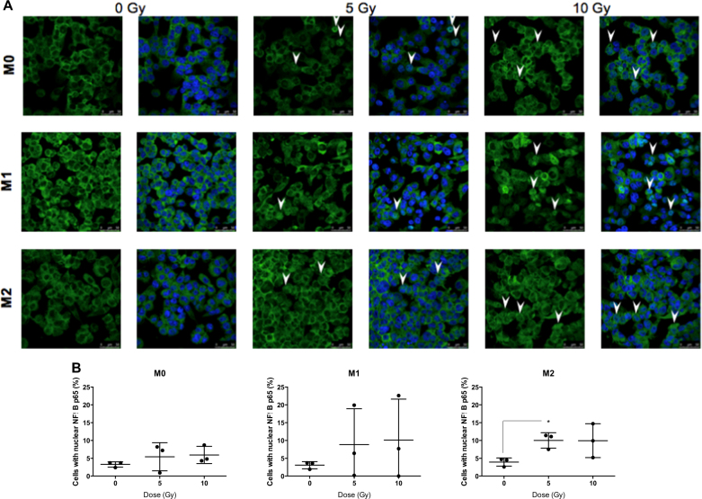
Fig. 5. Proton irradiation induces nuclear translocation of p65 (NFκB).
M0, M1, and M2 macrophages were irradiated by different doses of protons. 2 h after the irradiation, the nuclear translocation of p65 was evaluated by NFκB p65 immunofluorescence labeling. NFκB p65 is stained in green and nucleus appears in blue. a Nuclear translocation of NFκB p65 is indicated (arrow) on representative immunofluorescence labeling images for M0, M1, and M2 macrophages after irradiation. b Quantification of the mean NFκB p65 intensity per nucleus 2 h after proton irradiation. Results are expressed in percentage of cells with nuclear NFκB p65. Quantifications were performed on minimum five images per condition (N = 3, mean ± SD). An unpaired t test was performed on data (*p ≤ 0.05; **p < 0.01; ***p < 0.001)

