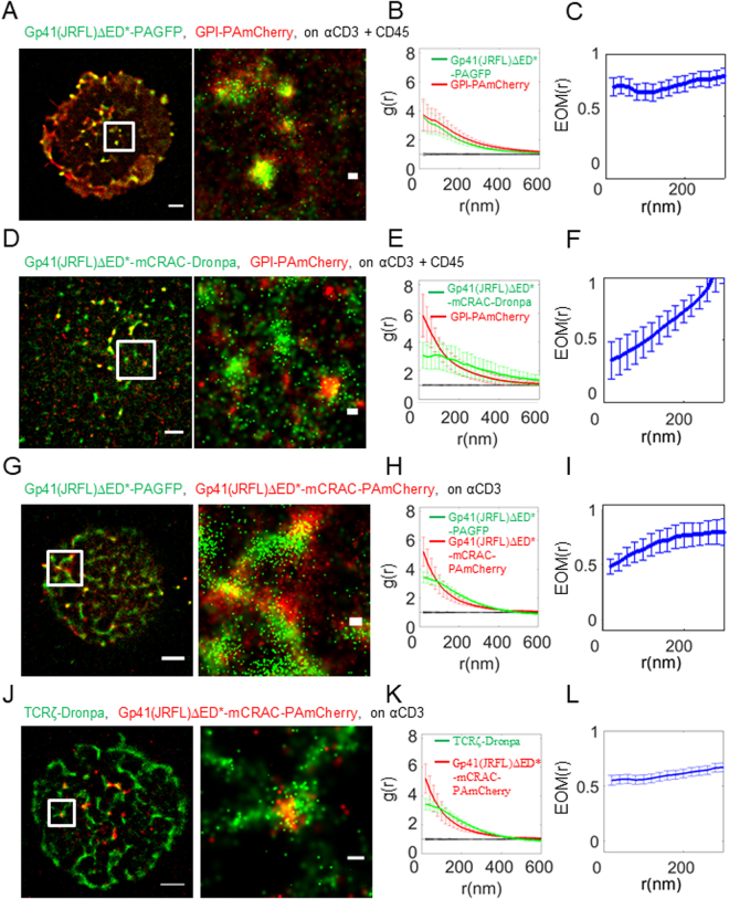
Figure 3.
Gp41-TCR interaction is not mediated by gp41 interactions with cholesterol. (A) Two-colour PALM imaging of fixed E6.1 Jurkat cells expressing gp41(JRFL)ΔED*-PAGFP and GPI-PAmCherry on coverslips coated with an αCD3 and αCD45. Cells were dropped and let spread on the coverslip for 3 min before fixation. Bars – 2 μm (left) and 200 nm (right). Shown is a representative cell (N = 16). (B) PCF of gp41-PAGFP (green) and GPI-PAmCherry (red). (C) EOM of gp41(JRFL)ΔED*-PAGFP and GPI-PAmCherry. (D) Two-colour PALM imaging of fixed E6.1 Jurkat cells expressing GPI-PAmCherry and gp41-mCRAC-Dronpa on coverslips coated with an αCD3 and αCD45. Cells were dropped and let spread on the coverslip for 3 min before fixation. (E) PCF of gp41(JRFL)ΔED*-mCRAC-Dronpa (green) and GPI-PAmCherry (red). (N = 6). (F) EOM of gp41(JRFL)ΔED*-mCRAC-Dronpa and GPI-PAmCherry. (G) Two-colour PALM imaging of fixed E6.1 Jurkat cells expressing gp41(JRFL)ΔED*-PAGFP and Gp41(JRFL)ΔED*-mCRAC-PAmCherry on an αCD3-coated coverslips. Cells were dropped and let spread on the coverslip for 3 min before fixation. (H) PCF of Gp41(JRFL)ΔED*-PAGFP (green) and gp41(JRFL)ΔED*-mCRAC-PAmCherry(red). (N = 7). (I) EOM of gp41(JRFL)ΔED*-PAGFP and gp41(JRFL)ΔED*-mCRAC-PAmCherry. (J) Two-colour PALM imaging of fixed E6.1 Jurkat cells expressing TCRζ-Dronpa and gp41(JRFL)ΔED*-mCRAC-PAmCherry on an αCD3-coated coverslips. Cells were dropped and let spread on the coverslip for 3 min before fixation. (K) PCF of TCRζ-Dronpa (green) and gp41(JRFL)ΔED*-mCRAC-PAmCherry(red). (N = 16). (L) EOM of TCRζ-Dronpa and gp41(JRFL)ΔED*-mCRAC-PAmCherry. Error bars are SEM.

