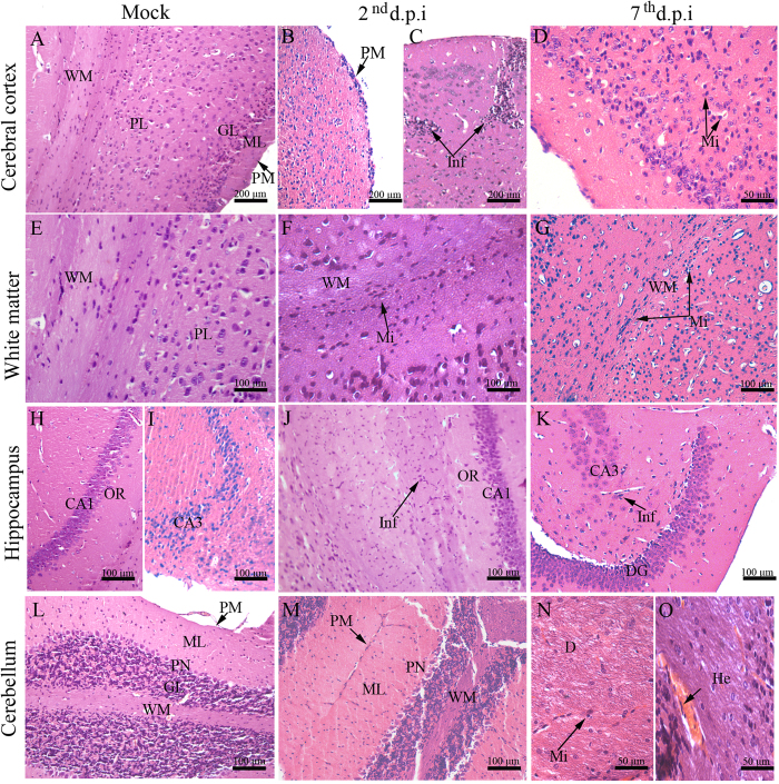
Figure 2.
Histopathological aspects of the brain and cerebellum tissues of DENV-infected mice. (A) Cerebral cortex of a mock-inoculated mouse exhibiting normal aspects; (B) Mouse infected with DENV-2 showing pia mater with inflammatory infiltrate; (C) focal perivascular inflammatory infiltrate on the 2nd d.p.i and (D) diffuse inflammatory infiltrate on the 7th d.p.i. (E) Normal white matter from a mock-inoculated mouse. (F) White matter committed with inflammatory infiltrate on the 2nd d.p.i and (G) on the 7th d.p.i. (H) CA1 and (I) CA3 hippocampal regions from a mock-inoculated mouse. (J) Microglial cell infiltrate in CA1 on the 2nd d.p.i and (K) in CA3 on the 7th d.p.i. (L) Cerebellum region with normal aspects extracted from a control mouse. (M) Degenerated Purkinje neuronal layer on the 2nd d.p.i. (N) Demyelination with microglial cell infiltrate and (O) hemorrhage on the 7th d.p.i. CA1 - Cornu ammonis 1 region; CA3 - Cornu ammonis 3 region; D - demyelination; DG - dentate gyrus; GL - granular layer; H - hippocampus; He - hemorrhage; ML - molecular layer; OR - orien; PL - pyramidal layer; PM - pia mater; Inf - Inflammatory infiltrate; PN - Purkinje neuron; Mi - Microglia; WM - white matter; d.p.i. - days post infection.

