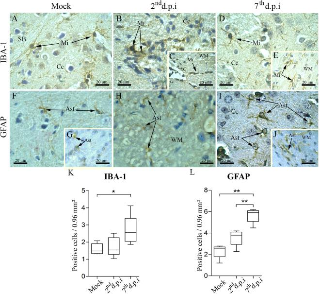
Figure 4.
Aspects of microglia and astrocytes in brain tissues of infected mice. Detection of microglia (IBA-1+ cells) in samples of the cerebral cortex and white matter from (A) non-infected, (B/C) 2nd d.p.i and (D/E) 7th d.p.i. (F/G) Detection of astrocytes (GFAP+ cells) in the cerebral cortex and white mater in samples from control animals. (H) Staining of astrocytes in the white matter of samples from infected mice on the 2nd d.p.i. (I/J) Staining of astrocytes in the cortex and in the white matter on the 7th d.p.i. Quantification of (K) IBA-1+ and (L) GFAP+ cells. Ast - astrocyte; C - capillary; Cc - cerebral cortex; Mi - microglia; WM - white matter; d.p.i. - days post infection. Statistical differences were evaluated using Mann-Whitney test (*p < 0.05; **p < 0.01).

