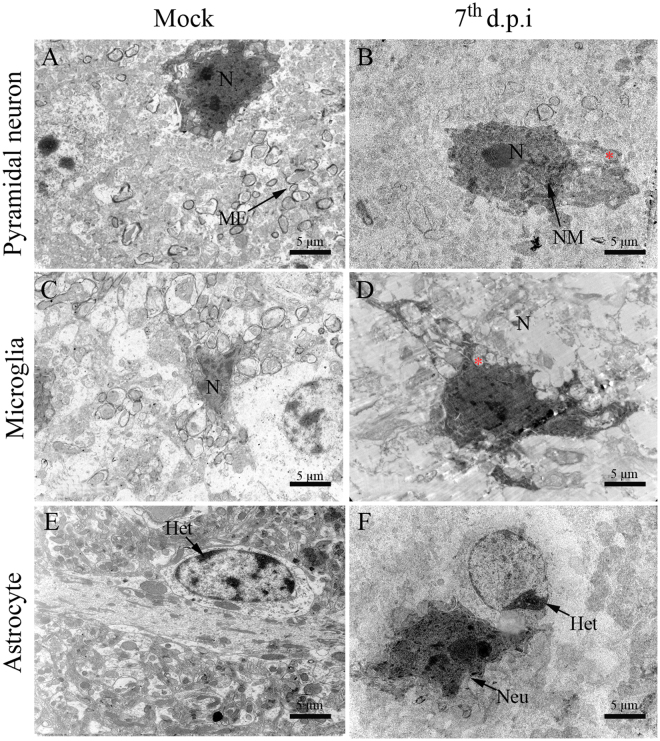
Figure 5.
Ultrastructural aspects of the brain tissue of BALB/c mice infected with DENV-2. (A) Pyramidal neurons, (C) microglial cells and (D) astrocyte of non-infected mice showing regular aspects. (B) Pyramidal neuron exhibiting increased nucleus, swollen mitochondria and irregular nuclear membrane. (D) Microglial cell with increased nucleus and swollen mitochondria. (F) Astrocyte with heterochromatin deposition. Samples from infected mice were considered on the 7th d.p.i. Het - heterochromatin; (Red asterisk) - mitochondria; MF - myelin fibers; N - nucleus; NM - nuclear membrane; d.p.i. - days post infection.

