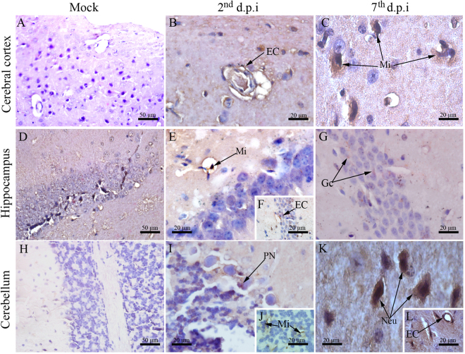
Figure 7.
Detection of DENV-NS3 protein in brain and cerebellum tissues. DENV-NS3 protein was detected using immunohistochemistry on brain and cerebellum cuts. Regions of (A) cerebral cortex, (D) hippocampus and (H) cerebellum from samples of control mice showing negative staining reaction for DENV-NS3. Samples from infected animals showing detection of DENV-NS3 in: (B) endothelial and (C) microglial cells located at the cerebral cortex on the 2nd and 7th d.p.i., respectively; (E,F) microglial/endothelial cells and (G) granular cells of the hippocampus on the 2nd and 7th d.p.i., respectively; (I,J) Purkinje neurons/microglial cells and (K,L) neurons/endothelial cells on the 2nd and 7th d.p.i., respectively. EC - endothelial cell; Mi - microglial cells; Gc - granular cells; PN - Purkinje neurons; Neu - neurons; d.p.i. - days post infection.

