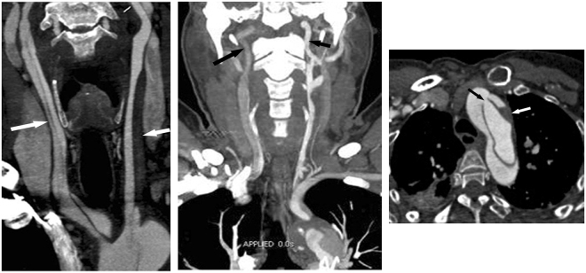
Figure 1.
Three CTA Risk Findings for Postoperative TND. (a) Carotid and aortic CTA in a 39-year-old man with ATAAD. Coronal thin MIP image showing aortic dissection extending to both the right (long arrow) and left CCA (short arrow). This finding suggests that dissection involving CCA is an important risk predictor for postoperative TND. (b) Carotid and aortic CTA in a 54-year-old man with ATAAD. Coronal thin MIP image showing that the contrast enhancement of the right ICA (long arrow) is lower than that of the left ICA (short arrow), due to the origin from the false lumen of the right CCA dissection. The results show an inadequate blood supply in the unilateral carotid artery and imply ipsilateral cerebral hypoperfusion. (c) Carotid and aortic CTA in a 49-year-old man with ATAAD. Axial image showing an entry tear located in the aortic arch (black arrow). Note the low density of thrombosis (white arrow) in the false lumen. An aortic arch tear implies an increased risk of pre- and intraoperative embolism into the cerebral artery via the entry point. CTA = CT angiography; TND = temporary neurological dysfunction; ATAAD = Acute type A aortic dissection; MIP = maximum intensity projection; ICA = internal carotid artery; MPR = multiplanar reformation. Note the low-density haematoma in the false lumen of the involved left CCA.

