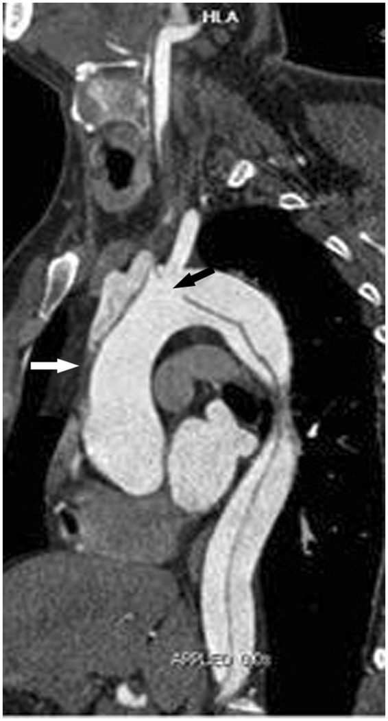
Figure 2.

Two CTA Risk Findings for Postoperative PND. Oblique sagittal MPR image showing dissection involving the thoracic aorta and supra-aortic branches. An entry tear (black arrow) is located in the aortic arch, and the intimal flap extends into the aAO in a retrograde fashion. Note the thrombosis (white arrow) in the false lumen, due to the integrated intimal flap in the aAO. The two CTA findings, which exist simultaneously, indicate an extremely high risk of thrombus in the false lumen extending to the cerebrum via the entry point. CTA = CT angiography; PND = permanent neurological dysfunction; MPR = multiplanar reformation; aAO = ascending aorta.
