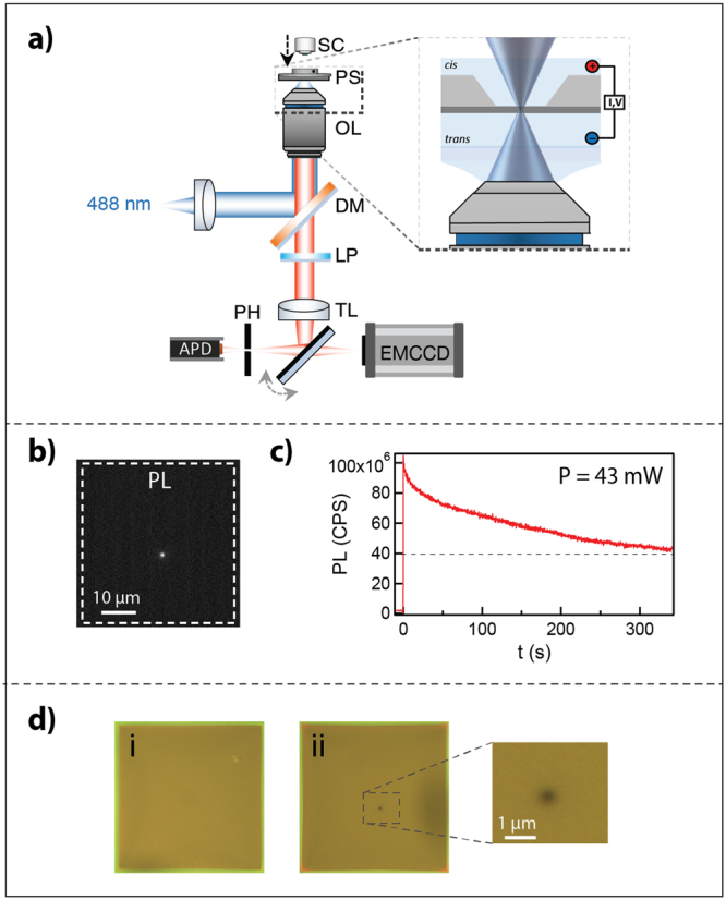
Figure 1.

Laser thinning of freestanding SiNx. (a) Schematic of the confocal setup. SC- SiNx chip; PS-piezo stage OL- objective lens; DM- dichroic mirror; LP- long pass filter; TL- tube lens PH- pinhole. The emission pathway is switchable between the APD and EMCCD. (b) Focusing of a ~45 mW 488 nm laser on the membrane results in photoluminescence emission, which is recorded by the APD in the >550 nm range. (c) Photoluminescence emission during laser-exposure, measured in counts per second. The laser is activated at t = 0 seconds. (d) Images of the 42 × 42 μm2 membrane under white-light illumination before etching (i). After 300 seconds of laser exposure, a thin region is visible as a contrasted spot (ii).
