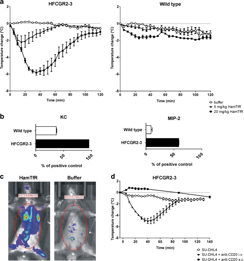
Fig. 2.
Humanized HFCGR2–3 mice display FIR upon mAb injection. (a), Change of body temperature of HFCGR2–3 (left panel) and wild type (right panel) mice injected with either buffer (open circles), 5 mg/kg (black triangles) or 20 mg/kg (black squares) of HamTfR antibody over a period of 2 h. (b), Serum levels of inflammatory cytokines KC (left panel) and MIP-2 (right panel) 2 h after infusion of 20 mg/kg HamTfR antibody in HFCGR2–3 and wild type mice. Serum levels are expressed as percentage of the assay’s internal positive control as described in Material and Methods and in Fig. S7. (c), In vivo production of ROS in HFCGR2–3 mice upon infusion of 20 mg/kg HamTfR antibody (left panel) or solvent buffer (right panel). (d), FIR as caused by infusing a mAb with different target specificity. Change of body temperature of HFCGR2–3 mice 2 h after injection of CD20-expressing human lymphoma cell line SU-DHL4 (black squares) or SU-DHL4 cells pre-incubated with human anti-CD20 antibody Rituximab injected i.v. (black triangles) or SU-DHL4 cells pre-incubated with Rituximab injected s.c. (open circles).

