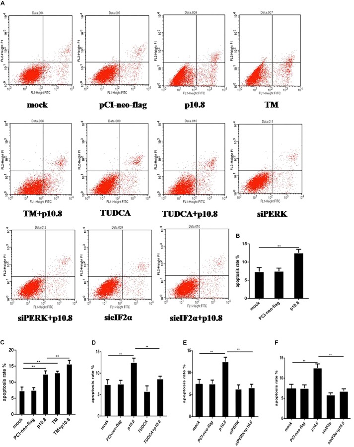
FIGURE 4.
Apoptosis was detected by flow cytometry. MDRV p10.8 protein-induced cell apoptosis was investigated in DF1 cells treated with (a) mock, (b) plasmid pCI-neo-flg, (c) recombinant plasmid pCI-neo-flg-p10.8, (d) 2 μg/mL TM, (e) pCI-neo-flg-p10.8+TM, (f) 2 μg/mL TUDCA, (g) pCI-neo-flg-p10.8+TUDCA, (h) siPERK, (i) pCI-neo-flg-p10.8+siPERK, (j) sieIF2α, (k) pCI-neo-flg-p10.8+sieIF2α, respectively. After 24 h, cells were stained with Annexin V-FITC/PI and the proportion of apoptotic cells in each group were detected by flow cytometry. (A) Images of apoptosis were determined via flow cytometry in DF1 cells. Statistical analysis of the proportions of cell apoptosis in (B) p10.8, (C) TM and pCI-neo-flg-p10.8+TM, (D) TUDCA and pCI-neo-flg-p10.8+TUDCA, (E) siPERK and pCI-neo-flg-p10.8+siPERK, (F) sieIF2α and pCI-neo-flg-p10.8+sieIF2α compared with mock and pCI-neo-flg. ∗P < 0.05, ∗∗P < 0.01, the same as in the following study.

