. 2018 Jun 15;10(4):406–419. doi: 10.4168/aair.2018.10.4.406

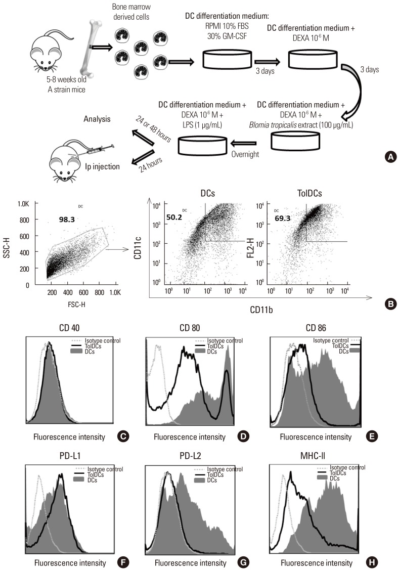
Fig. 1. Immunophenotype of DCs differentiated from bone marrow cells in the presence of dexamethasone. (A) Protocol for the generation of tolerogenic DCs from A strain mouse bone marrow. Control cells were cultured in differentiation medium and stimulated with LPS in the absence of dexamethasone. (B–H) Cells were stained with antibodies against surface markers as indicated. Debris and dead cells were excluded on the basis of forward-scatter and side-scatter. (B) Representative dot plots depicting the percentages of CD11c+CD11b+ cells. (C-H) Histograms of CD40 (C), CD80 (D), CD86 (E), PD-L1 (F), PD-L2 (G), and MHC II (H) cells gated on CD11c+CD11b+ double positive cells. DCs are represented by filled gray histograms and TolDCs by black lines; isotype controls are shown by dotted gray lines. Results are representative of 3 independent experiments. DC, dendritic cell; LPS, lipopolysaccharide; PD-L, programmed death-ligand; MHC, major histocompatibility complex; TolDC, tolerogenic dendritic cell.

Fig. 1