. 2018 Jun 15;10(4):406–419. doi: 10.4168/aair.2018.10.4.406

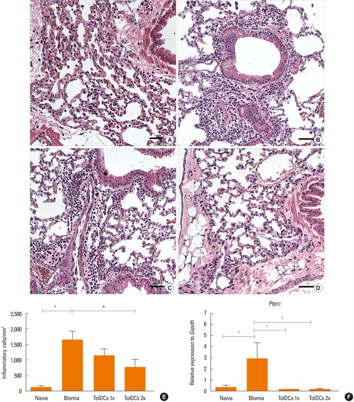
Fig. 5. Airway inflammation of TolDC-treated mice. The right lobe sections of the lungs were stained with H&E for the quantification of inflammatory cells by optical microscopy. For each of the lung 10 fields (400×) were analyzed per section, and the data used to calculate the mean number of cells per mm2. Scale bar, 50 µm. (A–D) Representative hematoxylin and eosin-stained sections of the lungs of A strain mice. (A) Normal tissue of an untreated animal. (B) Inflammatory infiltrate with a predominance of polymorphonuclear around bronchi and arterioles in an animal from the positive control group (Blomia). (C and D) Reduced lung perivascular infiltration by polymorphonuclear cells, in a Blomia-sensitized animal pre-treated with TolDCs 1× and TolDCs 2×, respectively. (E) Infiltration of inflammatory cells per mm2. (F) Relative expression to Gapdh of Ptprc. The data is representative of 3 independent experiments. TolDC, tolerogenic dendritic cell. *P<0.05; P<0.01; P<0.001 (Newman-Keuls multiple comparison test).

Fig. 5