. 2018 Jun 15;10(4):406–419. doi: 10.4168/aair.2018.10.4.406

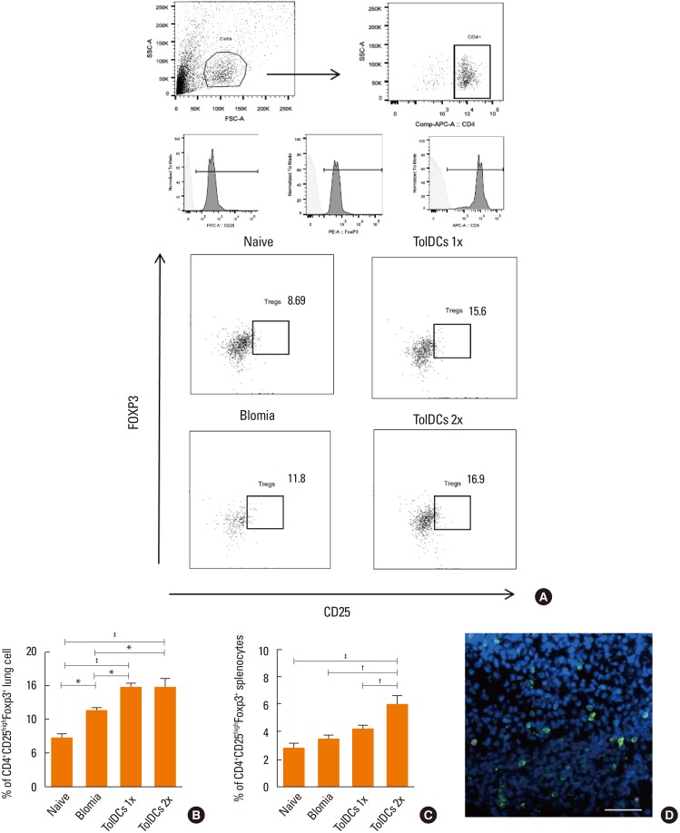
Fig. 7. TolDC treatment recruits Tregs to the lung and the spleen. The lung and spleen cell preparations obtained from mice of each experimental group were stained with anti-CD4-APC and anti-CD25-FITC, and after permeabilization the cells were stained with anti-Foxp3-PE. (A) Gating on CD4+ T cells, and representative dot plots of lung Tregs (CD4+CD25highFoxp3+) and isotype controls. (B) Quantification of lung Tregs (CD4+CD25highFoxp3+) relative to total CD4+ T cells. (C) CD4+CD25highFoxp3+ splenocytes. (D) Confocal microscopy images showing CD3 (green), Foxp3 (red), and nuclei stained with DAPI (blue) in the lung tissue of TolDC-treated mice. Values are expressed as means±SEM of 5–7 mice per group. TolDC, tolerogenic dendritic cell; Treg, regulatory T cell; FITC, fluorescein isothiocyanate; DAPI, 4′,6-Diamidino-2-phenylindole; SEM, standard error of the mean. *P<0.05; P<0.01; P<0.001 (Bonferroni's multiple comparison test).

Fig. 7