Fig. 1.
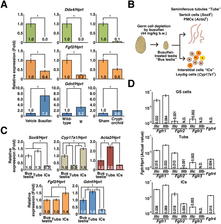
Identification of Fgf2-expressing cells and their targets in the testes. (A) RT-qPCR results: the effects of busulfan treatment (44 mg/kg body weight) on mice with a congenital spermatogenic defect (W mutation), and experimental cryptorchidism were analyzed. After normalization to Hprt expression level, the values of control groups (vehicle, sham, and wild-type groups for the busulfan, cryptorchid, and W groups, respectively) were set to 1.0 (n = 5–8 for each group). The testes were harvested at 4 weeks (busulfan treatment) or 7 weeks (experimental cryptorchidism) after treatment and subjected to analysis. W mouse testes were harvested at 8 weeks of age. Age-matched mice were used as control samples in each experiment. (B) Preparation of seminiferous tubules and testicular interstitium. The testes were harvested at 4 weeks after treatment with 44 mg/kg body weight busulfan (Bus testes) and subjected to collagenase digestion to separate interstitial cells (ICs) from the seminiferous tubules (Tubs). The resulting populations were subjected to RT-qPCR. (C) Gene expression of niche components. After normalization to Hprt expression level, the value of the “Bus testis” group was set to 1.0 (n = 6 for each group). (D) Fgfr expression in GS cells and niche components. The value of each sample is indicated after normalization to Hprt expression (n = 3 independent cultures of GS cells and n = 6 for Tubs and ICs). Results are shown as the mean ± SEM. Daggers (†) indicate statistically significant differences between treatment groups (P < 0.05).

