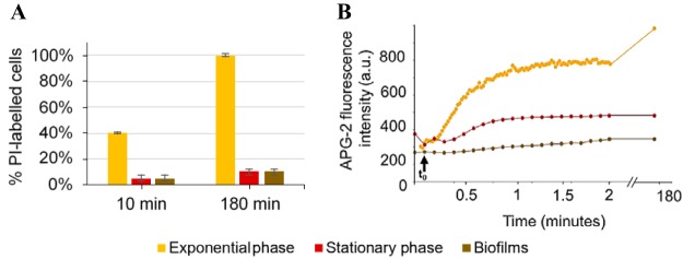
FIG 4.
PI uptake and potassium ion release in response to daptomycin treatment. Significant membrane damage only occurs within exponentially growing S. aureus cells. (A) Percentage of PI-labeled bacterial cells 10 min and 180 min after daptomycin addition. Error bars represent the standard deviation, based on at least 5 independent acquired images. (B) APG-2 fluorescence intensity evolution over time after daptomycin addition. Bacteria were incubated for 10 min with APG-2 (a membrane-impermeant K+ ion marker) to check the fluorescence intensity signal. t0 corresponds to daptomycin addition (indicated by black arrow).

