
FIG 4.
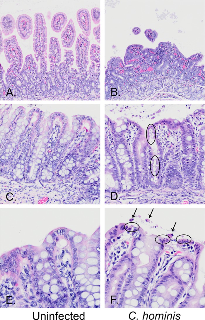
Hematoxylin- and eosin-stained sections from the spiral colons of uninfected (A, C, and E) and C. hominis-infected (B, D, and F) piglets, magnified ×20 (A to D) and ×40 (E and F) and showing normal structures (left panels) compared to representative lesions from C. hominis-infected pigs (right panels). (A and B) Normal duodenal villi within an uninfected pig (A) compared to marked villus blunting and fusion and moderate lymphocytic infiltrates in the lamina propria of a C. hominis-infected pig (B). (C and D) Normal spiral colon surface and glandular epithelia (C) compared to a gland heavily colonized with C. hominis, with epithelial cell loss, attenuation, and sloughing into the lumen (D). (E and F) Normal spiral colon surface (E) compared to a surface mildly colonized by C. hominis, with single-cell necrosis and sloughing (F). The arrows mark apoptotic epithelial cells sloughing into the lumen. The ovals encircle epithelial cells that are infected with C. hominis organisms at the very apical surface of the epithelial cells.