. 2016 May 13;7(8):5523–5529. doi: 10.1039/c6sc01279e

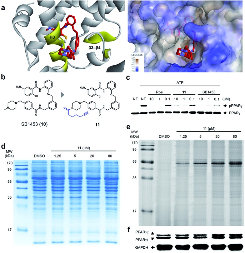
Fig. 6. (a) X-ray co-crystal structure of the SB1453–PPARγ LBD reveals the exposure of the piperidine moiety at the entrance of the PPARγ binding pocket. (b) Structure-guided rational design of the target identification probe 11 to demonstrate the target selectivity. (c) In vitro Cdk5 assay of PPARγ on treatment with rosiglitazone, 11, or SB1453 in a dose-dependent manner. (d, e) Coomassie staining and fluorescence scanning patterns of SDS-PAGE of lysates from differentiated 3T3-L1 adipose cells treated with probe 11 in various concentrations. (f) Western blot against the same gel in (e) to confirm PPARγ as the target proteins labeled by 11.

Fig. 6