. 2016 Jun 13;7(9):5624–5634. doi: 10.1039/c6sc00523c

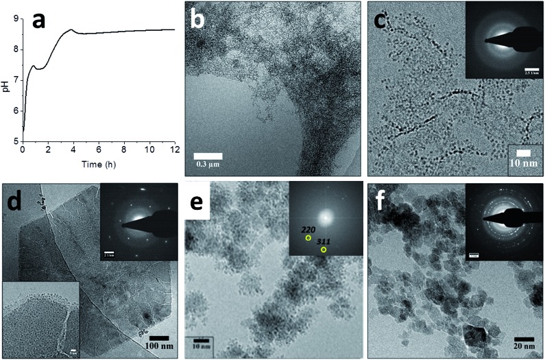
Fig. 6. Magnetite formation from a ferrihydrite precursor phase by ammonia diffusion. (a) pH curve through time for magnetite synthesis from ferrihydrite and Fe2+ upon in-diffusion of NH3, showing two distinct nucleation events. (b–f) Cryo-TEM images of (b) the ferrihydrite precursor, showing its gel-like character, (c) ferrihydrite stabilized by Fe2+ at pH ∼ 5, showing the 1.5–2.0 nm primary particles (inset: SAED pattern, showing no crystallinity), (d) the hexagonal green rust intermediate after ∼1 hour reaction time (insets: SAED pattern with 3.9 nm–1 spacings and higher magnification of a corner of the crystal, showing the 1.0–1.5 nm secondary particles), (e) the formation of magnetite through the aggregation of secondary particles (inset: FFT pattern showing lattice spacings of magnetite, while the contours of the secondary particles are still clearly visible in the aggregates), and (f) the final magnetite product (inset: SAED pattern, displaying the common magnetite reflections). Please note that the cryoTEM image in (e) was recorded in a solution with higher Fe2+ concentration than the other images. Reproduced from ref. 91.

Fig. 6