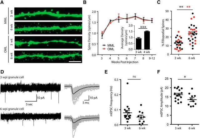
Figure 2.
Synaptic maturation and development. A, Spine density measurements in MML (top) and OML (bottom) of 3-week-old and 6-week-old dentate granule cells. Scale bar, 5 μm. B, Developmental increase in spine density across cell development in MML (black) and OML (red). Inset, Average spine density across layers increases between 3 and 4 weeks after mitosis, then remains constant between 4 and 12 weeks. C, Developmental increase in the percentage of spines with filopodial morphology in both MML (black) and OML (red). D, mEPSC recordings in the presence of 10 μm SR95531 and 1 μm TTX to isolate excitatory events. mEPSCs were recorded at 3 and 6 weeks after mitosis. E, There was no difference in the mEPSC frequency at 3 and 6 weeks after mitosis. F, There was a significant decrease in mEPSC amplitude, suggesting weaker synaptic inputs at 6 weeks after mitosis. *p < .05, **p < .01, ***p < .001.

