
Figure 3.
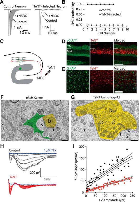
Silencing synaptic input with tetanus toxin expression. A, Comparison of synaptic responses in autaptically cultured neurons in control (left) and following tetanus toxin infection (TeNT). The shaded area indicates the AMPA-receptor EPSC. B, Expression of TeNT abolishes synaptic responses in autaptically cultured neurons. C, Schematic of TeNT viral injection into the medial entorhinal cortex, which will functionally silence axons in the MML of the dentate gyrus. D, Expression of TeNT in the MML dramatically reduces the intensity of VGluT1 expression in the MML, indicating a disruption of presynaptic function. Scale bar, 100 μm. E, TeNT expression did not elicit astrogliosis. F, G, Electron micrographs from control (F) and TeNT-overexpressing (G) axons. TeNT expression results in axonal swelling and vesicle accumulation (b, axonal bouton; s, dendritic spine; arrowheads, synapses). Scale bar, 500 nm. H, fEPSP recordings from the MML while electrically stimulating the MPP fibers. I, fEPSP responses were significantly attenuated when TeNT was expressed in the MML, without changing fiber volley amplitudes.