Figure 2.
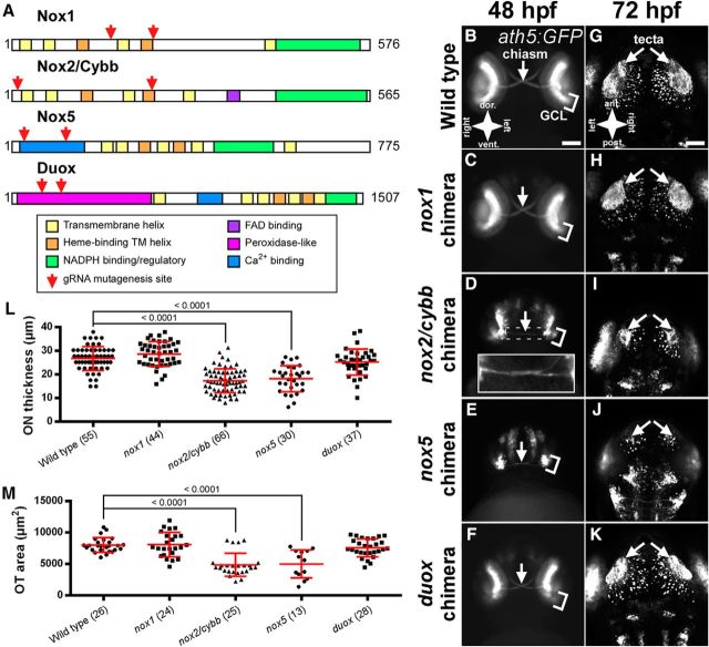
Chimeric nox mutants show optic nerve and tectal defects. A, Schematic of zebrafish NOX protein domains as determined by alignment to the human protein sequences. Red arrows indicate the relative location of gRNA target sites. B–F, Anterior views Tg(ath5:GFP)-positive, nox chimeras at 48 hpf. White arrows indicate the optic chiasm. Inset in D shows in enlarged and enhanced view of the optic chiasm. G–K, Dorsal views of chimeric embryos showing the innervation of the optic tecta (white arrows). GFP-positive cells not indicated with arrows represent nontectal early neurons from the hindbrain and eye. Scale bars: B–F, 100 μm; G–K, 50 μm). L, Graph of ON thickness in Tg(ath5:GFP) positive, chimeric mutants. M, Graph of OT area in Tg(ath5:GFP)-positive, chimeric mutants. Numbers in parentheses indicate the number of embryos analyzed. Graphs show mean ± SD. p-values from Dunnett's tests are reported above comparison bars.

