Figure 5.

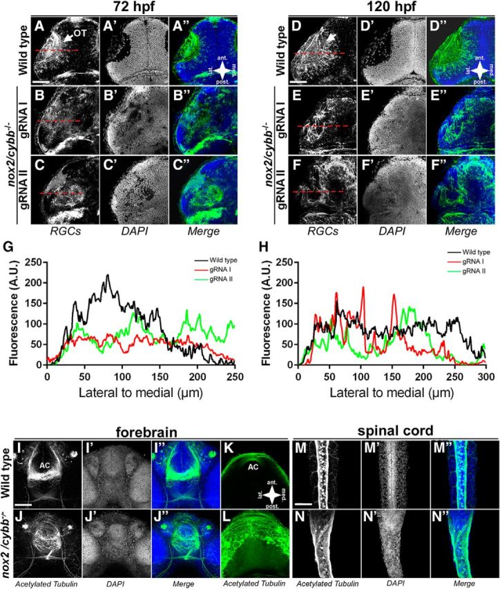
Axons are mistargeted in the OT of homozygous nox2/cybb mutants. A–C′′, Micrographs of 72 hpf WT (A) and nox2/cybb−/− (B, C) midbrains labeled with zn-8 (A–C) and DAPI (A′–C′). Mutants show defects in tectal innervation. D–F′′, WT (D) and nox2/cybb−/− midbrains at 120 hpf labeled with zn-8 (E, F) and DAPI (E′–F′) showing that the phenotype does not recover at later stages. White arrows indicate the optic tecta. G, H, Line scans plotting fluorescent intensity of zn-8 signals versus position in the midbrain. Placements of the line scans are shown with red dashed lines in zn-8 panels. I–L, Micrographs of 48 hpf WT (I–K) and nox2/cybb−/− (J–L) forebrains labeled with anti-acetylated α-tubulin antibody and DAPI. nox2/cybb−/− embryos exhibit aberrant axonal projections at the forebrain AC. Ventral views are shown in I and J; dorsal views in K and L. M–N″, Micrographs of 48 hpf WT (M–M″) and nox2/cybb−/− (N–N″) spinal cord labeled with anti-acetylated α-tubulin antibody and DAPI. The spinal cord is wider and shows aberrant projections of the dorsal longitudinal fascicle in nox2/cybb−/− embryos (N–N″) compared with WT embryos (M–M″). Scale bars, 100 μm.
