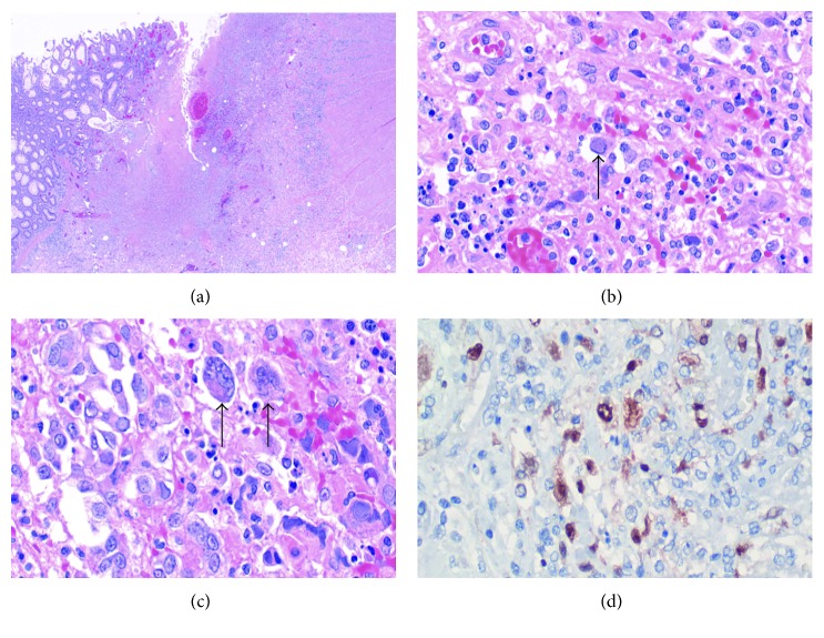
Figure 1.
(a) Section from the ulcerated colonic mucosa with deep fissuring ulcer. Viral cytopathic changes are noted. (b, c) Giant cells within ulcer with characteristic intranuclear Cowdry type A inclusions of HSV-1 and HSV-2. Black arrows point to individual giant cells with Cowdry type A inclusions. (d) Immunohistochemistry study for HSV-2 antibody highlighted by the brown nuclear staining.

