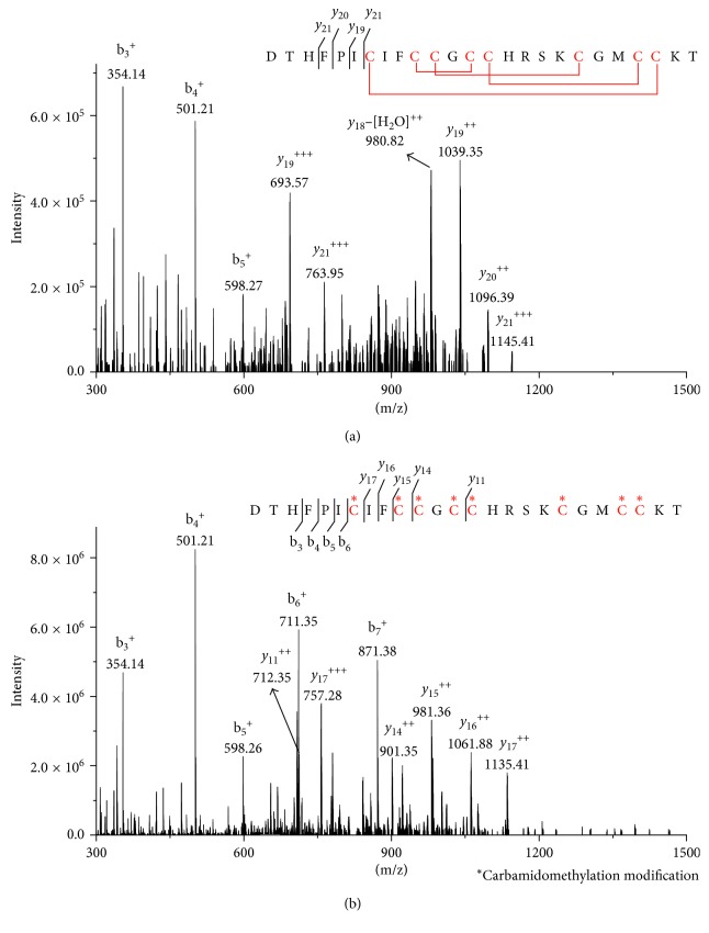
Figure 1.
MS/MS spectrum of hepcidin-I and hepcidin-M. (a) is the MS2 spectrum for 558.6 m/z (z = 5) derived from the hepcidin-I, and (b) is for 651.8 m/z (z = 5) derived from the hepcidin-M. The cysteine (C) amino acid sequence of red color indicates disulfide bond and carbamidomethylation position in (a) and (b).

