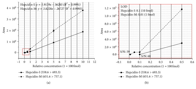
Figure 3.
The graph shows the peak area for hepcidin amount ranging from 1 fmol to 1 pmol (1, 5, 10, 50, 100, and 500 fmol and 1 pmol). The graph of hepcidin-I and -M were presented using 558.6 > 693.3 and 651.6 > 757.1 of transitions, respectively. (a) R2 of hepcidin-I and -M were obtained by the equations of y = 1.9139x−56285 and y = 3.8228x−26727, respectively. (b) An enlarged graph of the red box portion in (a), showing the peak area according to the amount of 1 fmol to 50 fmol and S/N value of LOD for both hepcidin-I and hepcidin-M.

