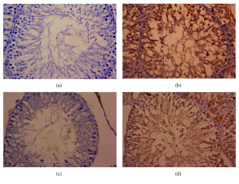
Figure 10.
Testicular expression of Bax protein was detected using immunohistochemical staining in (a) control, (b) cisplatin (CDDP), (c) olive leaf extract (OLE), and (d) OLE + CDDP groups. In the control and OLE groups, Bax-positive brown-stained cells were sparse and weakly immunostained. However, many testicular cells exhibited apoptosis and were stained brown (Bax positive) due to CDDP. In the OLE + CDDP group, the number of Bax-positive cells was markedly increased. (400x).

