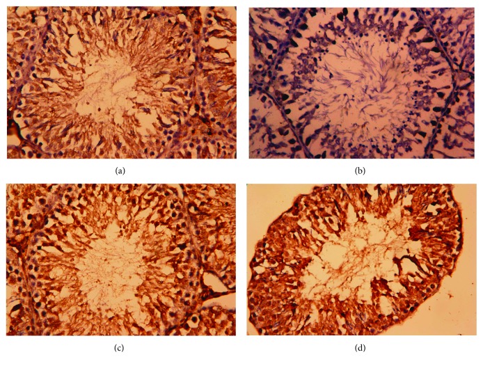
Figure 11.
Testicular expression of proliferating cell nuclear antigen (PCNA) protein was detected using immunohistochemical staining in (a) control, (b) cisplatin (CDDP), (c) olive leaf extract (OLE), and (d) OLE + CDDP groups. In the control and OLE groups, PCNA-positive brown-stained cells were moderately to strongly immunostained. However, many testicular cells were weakly stained with brown color due to CDDP. In the OLE + CDDP group, the number of PCNA-positive cells was markedly increased. (400x).

