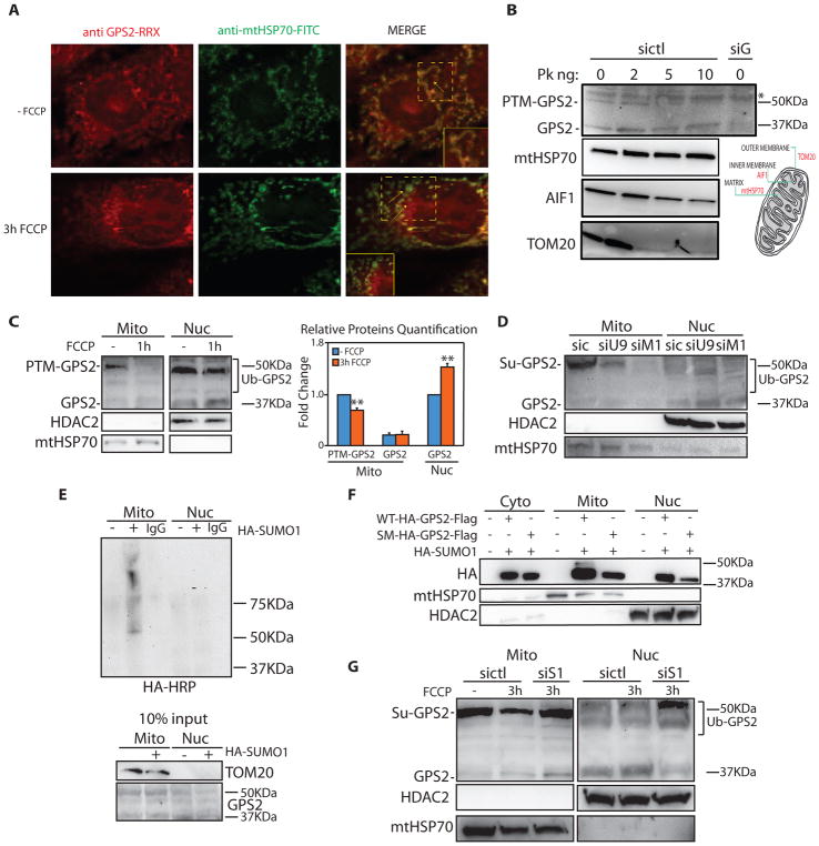
Figure 4. Mitochondria-to-nucleus regulated translocation of GPS2 in response to mitochondria depolarization.
(A) Reduced GPS2 localization to the mitochondria and corresponding increased nuclear staining (see inset and arrows) upon 3h of FCCP in 3T3-L1 cells.
(B) Proteinase K protection assay. Non-modified GPS2 and post-translationally modified GPS2 (PTM-GPS2) localize respectively to the matrix and OMM. Degradation patterns of TOM20, AIF1 and mtHSP70 are representative of proteins in the OMM, IMM and matrix.
(C) WB of fractionated extracts showing mitochondria-to-nucleus translocation of GPS2 in 3T3-L1 cells upon 1h FCCP (relative quantification on the right).
(D) GPS2-PTM is reduced by downregulation of either Ubc9 (siU9) or Mul1 (siM1).
(E) Sumoylation assay with HA-SUMO1 in 293T cells.
(F) Mitochondria localization of HA-GPS2-Flag WT or Sumo-mutant (SM: K45, K71R) in 293T cells.
(G) Impaired translocation of GPS2 in FCCP-treated cells transfected with siSENP1 (siS1).

