
Fig. 1.
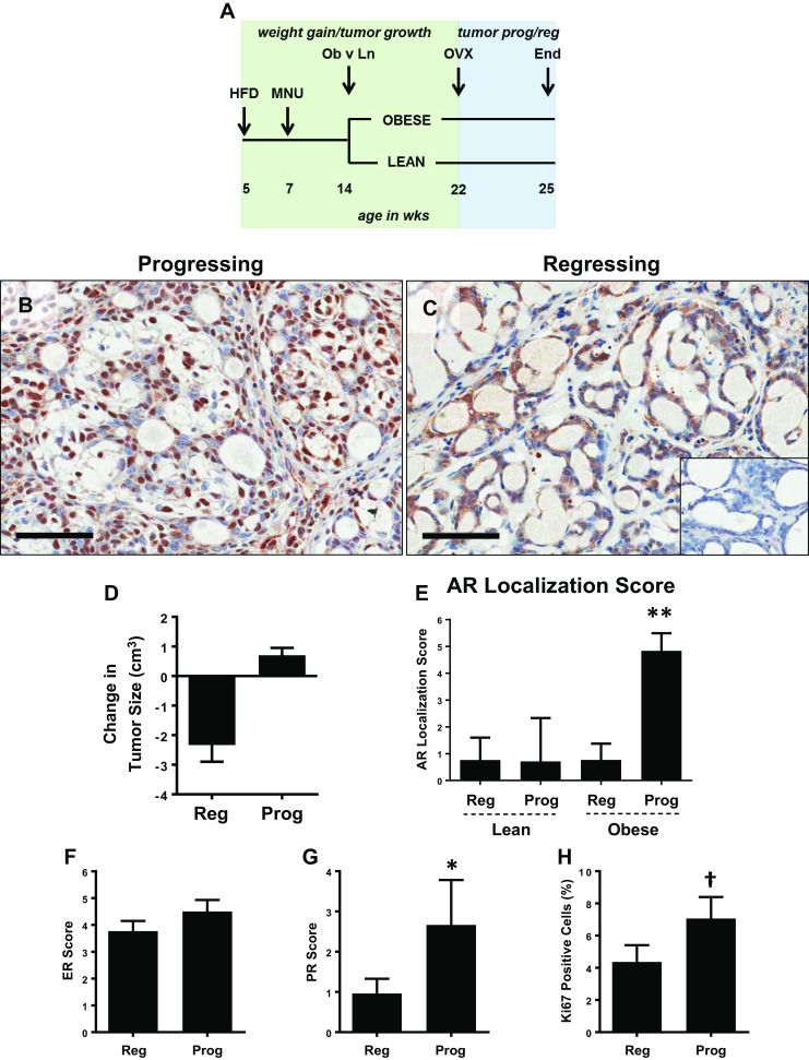
AR is localized to the nucleus in progressing tumors from obese rats. a Schematic study design. HFD high fat diet, MNU N-methyl-N-nitrosourea, OVX ovariectomy. b–c Representative images of AR immunostaining showing b nuclear and c cytoplasmic localization. Scale bars = 60 μm. Inset in c shows an AR negative (0% cells positive) tumor. d Change in size of regressing (shrinking) and progressing (growing) tumors from OVX until the end of study (3 weeks post OVX) in lean and obese rats. Regressing were defined as those with a size change <0 cm3, and progressing were defined as those with a size change >0 cm3, measured from the time of OVX until the end of the study (3 weeks post OVX). e AR localization score, calculated as Nuclear AR Score – Cytoplasmic AR Score in regressing (reg) and progressing (prog) tumors from lean and obese rats. The maximum cytoplasmic or nuclear AR score is 12, so the AR localization score can range from −12 (strong AR intensity, exclusively cytoplasmic) to 12 (strong AR intensity, exclusively nuclear). T test determined statistical significance. N = 17 regressing and 7 progressing tumors from lean, and 26 regressing and 6 progressing tumors from obese rats. **p < 0.01 f–h IHC scores for ER (f), PR (g) based on the Allred system, and percent Ki67 positive cells (h) in tumors from obese rats, categorized as regressing (reg) or progressing (prog). Mann-Whitney test determined statistical significance. N = 26 regressing and 6 progressing tumors. *p < 0.05; †p < 0.1