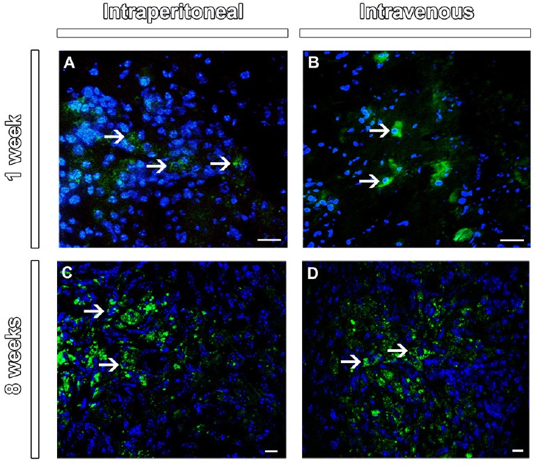
Figure 1.

Tracking of mesenchymal stem cells in the lesion epicenter of the spinal cord at 1 and 8 weeks after injection.
Merge images with co-localization of GFP+ (green) and DAPI+ (blue, arrows) MSC at the lesion epicenter in the cross sections of spinal cord at 1 week (A and B) and 8 weeks (C and D) after injection. Scale bars: 20 µm (A and B) and 10 µm (C and D). Sections were analyzed under confocal microscopy (Disk scanning unit). GFP: Green fluorescent protein; MSC: mesenchymal stem cells; DAPI: 4′,6-diamidino-2-phenylindole.
