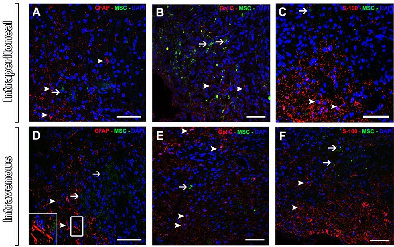
Figure 2.

Expression of glial cell markers in the injured spinal cord at 8 weeks after MSC injection.
The lack of co-localization at merge images for GFAP (A and D), Gal-C (B and E) and S100 (C and F). Arrows indicate GFP+ MSC (green) and arrowheads indicate glial markers (red). DAPI nuclear label is in blue. Note the star-shaped GFAP staining in the figure D insert. Scale bars: 50 µm. Sections were analyzed under confocal microscopy (disk scanning unit). GFAP: Glial fibrillary acidic protein; GFP: green fluorescent protein; MSC: mesenchymal stem cells.
