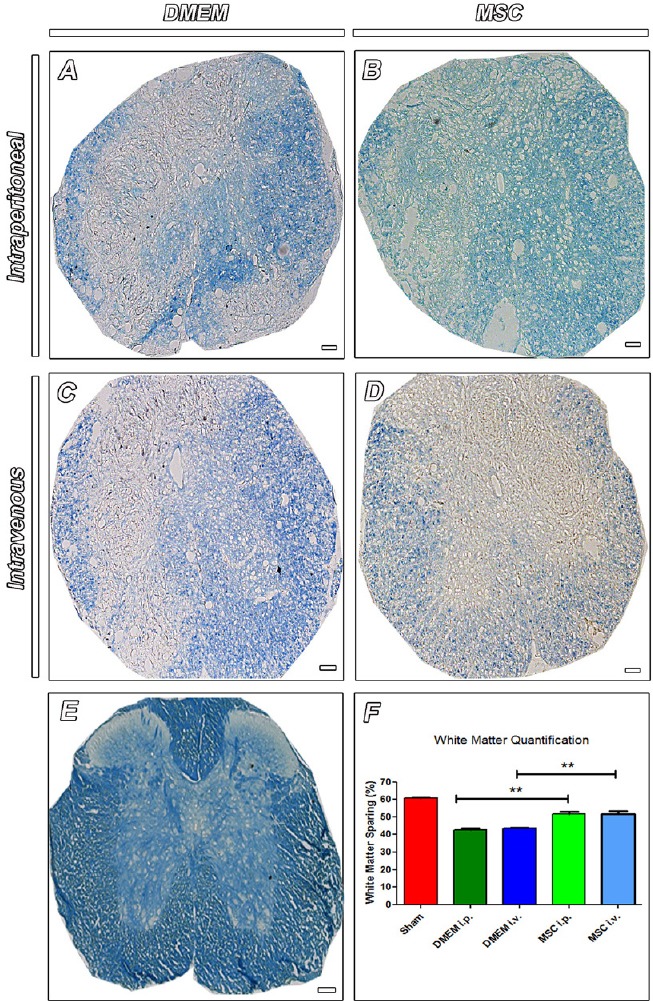
Figure 4.

White matter sparing in the injured spinal cord at 8 weeks after cell transplantation.
(A–D) Cross sections of spinal cord stained with luxol fast blue (LFB), a stain commonly used to observe myelin under light microscopy. Note tissue disorganization and less-intensely stained area in animals injected with DMEM (A – DMEM i.p. and C – DMEM i.v.) compared to animals that received MSCs (B – MSC i.p. and D – MSC i.v.). Staining with LFB also revealed the normal white matter distribution in sham-operated animals (E). Scale bars: 50 µm. Quantitative analysis showed higher values for areas of spared white matter for MSC-treated animals compared to the DMEM-treated ones (F). n = 3 per group. Results are expressed as the mean ± SEM (**P < 0.01). Sections were analyzed under optic microscope (Axioscop 2 Plus - Zeiss). One-way analysis of variance followed by Tukey's post hoc test was used. MSC: Mesenchymal stem cells; DMEM: Dulbecco's Modified Eagle's medium.
