Figure 5.

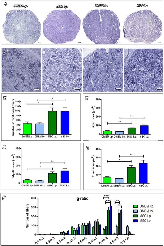
Morphometry of myelinated nerve fibers in toluidine blue-stained semithin sections of the injured spinal cord.
The increased tissue disorganization in groups that received only the injection of DMEM compared to groups that received the injection of MSCs (A and A’). Scale bars: 20 µm. A larger number of myelinated fibers are observed in animals that received transplants of cells (B). MSC-transplanted animals also showed larger area of axons (C), myelin (D) and fiber (E). In F, MSC animals had more fibers in the optimal range for spinal cord g-ratio, which is correlated with better conduction velocity (0.7–0.8 range). n = 3 per group. Results were expressed as the mean ± SEM. *P < 0.05, **P < 0.01, ***P < 0.001. Sections were analyzed under optic microscope (Axioscop 2 Plus - Zeiss). One-way analysis of variance followed by Tukey's post hoc test was used. DMEM: Dulbecco's Modified Eagle's medium; MSC: Mesenchymal stem cells.
