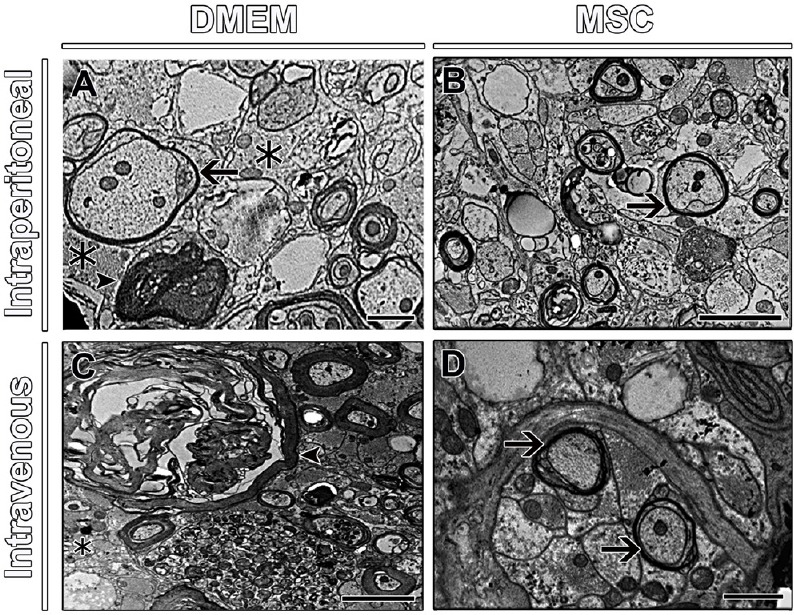
Figure 6.

Ultrastructural observation of spinal cord parenchyma of mice.
The presence of astrocyte processes (asterisks) and degeneration fibers (arrowhead) in groups that received DMEM (A, C), while groups that received MSCs show regenerating axon sprouts with evidence of myelination (arrows) (B, D). Arrow in A indicates an axon being remyelinated. Scale bars: 1 µm. Sections were analyzed under transmission electron microscope (Zeiss 900 Transmission Electron Microscope - Zeiss). DMEM: Dulbecco's Modified Eagle's medium.
