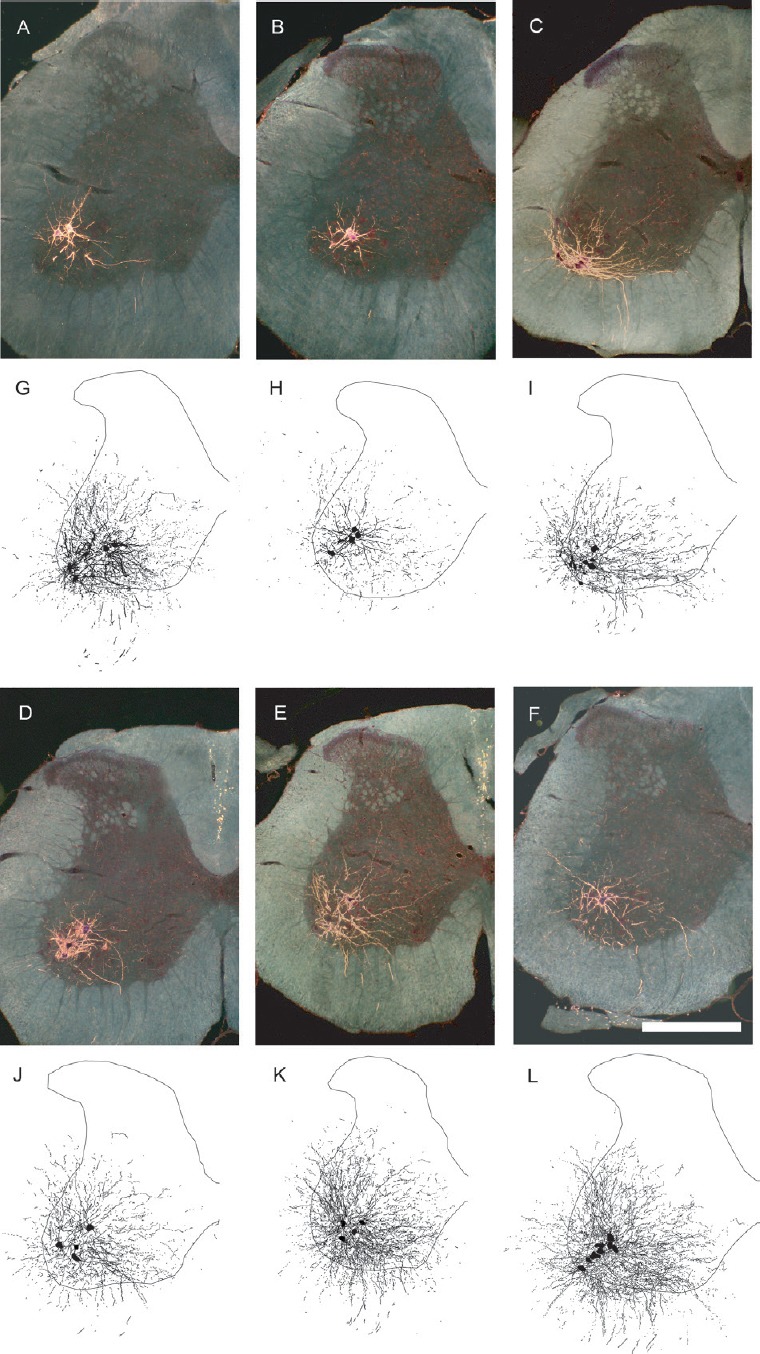
Figure 1.

Motoneuron morphology is protected by gonadal hormones following spinal cord injury.
Darkfield digital micrographs and matching computer-generated composites of transverse hemisections through the lumbar spinal cords of a sham animal (A, G), an injured animal given a blank implant (SCI; B, H), an estradiol-treated injured animal (SCI + E; C, I), a dihydrotestosterone-treated injured animal (SCI + D; D, J), an injured animal treated with both hormones (SCI + E + D; E, K), and a testosterone-treated injured animal (SCI + T; F, L), after horseradish peroxidase conjugated to the cholera toxin B subunit (BHRP) injection into the left vastus lateralis muscle. Computer-generated composites of BHRP-labeled somata and processes were drawn at 480 μm intervals through the entire rostrocaudal extent of the quadriceps motor pool; these composites were selected because they are representative of their respective group average dendritic lengths. Scale bar: 500 µm. (Images from Byers et al. (2012) and Sengelaub et al. (2018).
