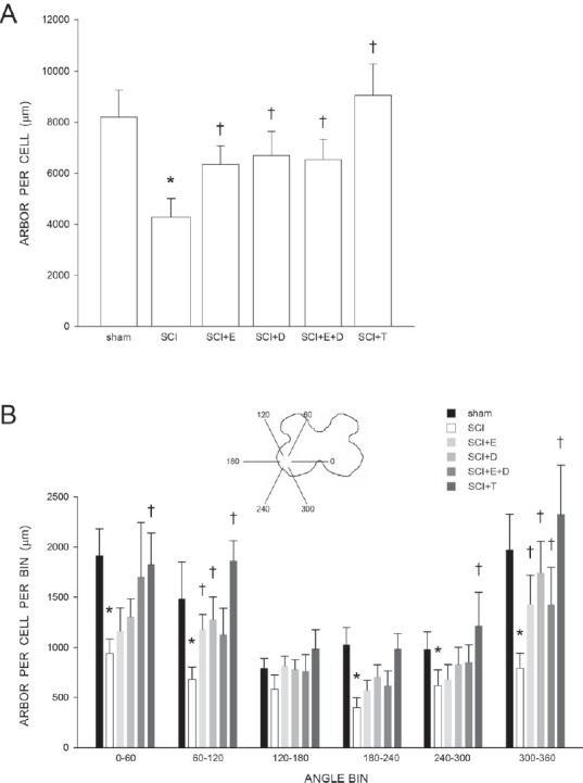
Figure 2.

Motoneuron dendritic length and distribution is protected by gonadal hormones following spinal cord injury.
(A) Dendritic lengths of quadriceps motoneurons of sham animals and injured animals that were either untreated (SCI), or treated with estradiol (SCI + E), dihydrotestosterone (SCI + DHT), estradiol and dihydrotestosterone combined (SCI + E + DHT), or testosterone (SCI + T). Following contusion injury, surviving quadriceps motoneurons lost over 50% of their dendritic length. Treatment with hormones attenuated this dendritic atrophy. (B) Inset: Drawing of spinal gray matter divided into radial sectors for measure of quadriceps motoneuron dendritic distribution. Quadriceps motoneuron dendritic arbors normally display a non-uniform distribution, with the majority of the arbor located between 300° and 120°. Following contusion injury, surviving quadriceps motoneurons in untreated animals (SCI) had reduced dendritic lengths throughout the radial distribution, especially ventromedially (60%, 300° to 360°). Treatment with hormones attenuated these reductions. Bar heights represent the mean ± SEM. *indicates significantly different from sham animals, † indicates significantly different from untreated SCI. (Data from Byers et al. (2012) and Sengelaub et al. (2018).
