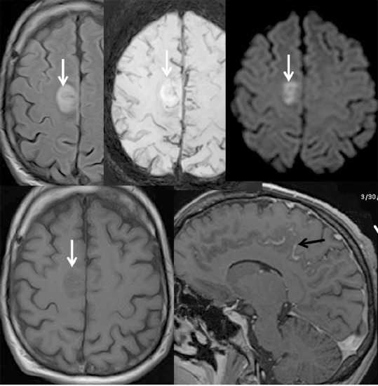
Figure 3.

Brain MRI of a 30-year-old male with acute myeloid leukemia who received allogeneic hematopoietic cell transplantation on December 19, 2017.
Right frontal cortical parasagital lesion with “acute” pattern (restriction of water on diffusion weighted imaging (DWI))- (white arrow) and leptomeningeal enhancement (black arrows).
