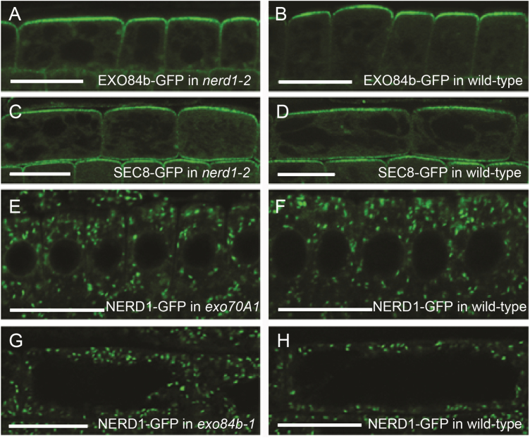
Fig. 3.
Subcellular localizations of NERD1 and exocyst markers are independent of each other. The localization of exocyst markers to the outer surface of root epidermal cells of nerd1-2 mutants (A, C) is similar to that in wild-type siblings (B, D). Conversely, the predominant localization of NERD1–GFP in the cytoplasm of root epidermal cells of exocyst mutants (E, G) is similar to that of wild-type siblings (F, H). Shown are epidermal cells in the root transition zone (A–D, G, H) and meristem (E, F). Confocal images provide radial longitudinal sections through the center of the root (A, B, E, F) in which the upper portion of cells shown are on the root surface. In tangential sections (C, D, G, H) parallel with the root surface the lateral walls of the epidermal cells are shown. Scale bars: 20 µm. (This figure is available in colour at JXB online.)

